 Orvos Image Evaluation Application
Confident retinal grading through independent, double-read evaluation.
Orvos Image Evaluation streamlines fundus image review — upload patient images and data, route them to a reading-doctor group for independent grading, automatically flag disagreements for adjudication, and export audit-ready reports.
Overview
A power user uploads fundus images and a spreadsheet of patient information — either in bulk or one patient at a time. Each patient is placed into a doctor group and graded independently by two reading doctors, Doctor A and Doctor B, who record retinal conditions for the right (OD) and left (OS) eyes.
When both reads agree, the patient is automatically marked successful. When they disagree, the patient is escalated to an evaluation doctor, Doctor C, who reviews both prior assessments and records the final diagnosis. At any point, power users and admins can monitor pending work and export a complete, status-tracked report.
Evaluation workflow
Independent reads with automatic adjudication when reading doctors disagree.
Upload
Power user uploads eye images and an Excel file of patient information.
Independent reads
Doctor A and Doctor B independently evaluate left and right eye images for retinal conditions.
Agreement check
If both doctors agree independently, the evaluation is marked successful.
Adjudication
If they disagree, Doctor C reviews both assessments and selects the final diagnosis.
Exportable reporting
A report is generated and exportable via Excel, showing patient information and the status of each evaluation.
Profiles & access
Users have secure accounts with role-based profiles.
Admin
All functions, including creating user accounts.
Power User
Upload images and Excel files, and run reports.
Reading Doctor
Evaluate images and record retinal conditions.
Evaluation Doctor
Review anomalies flagged by reading doctors and adjudicate.
Selectable conditions
Standardized diagnostic options available to evaluating doctors.
Product screens
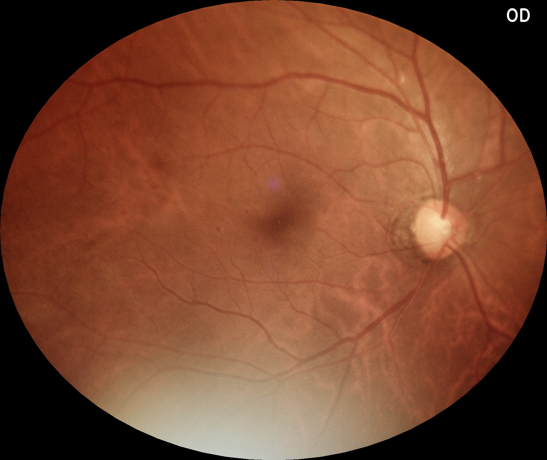
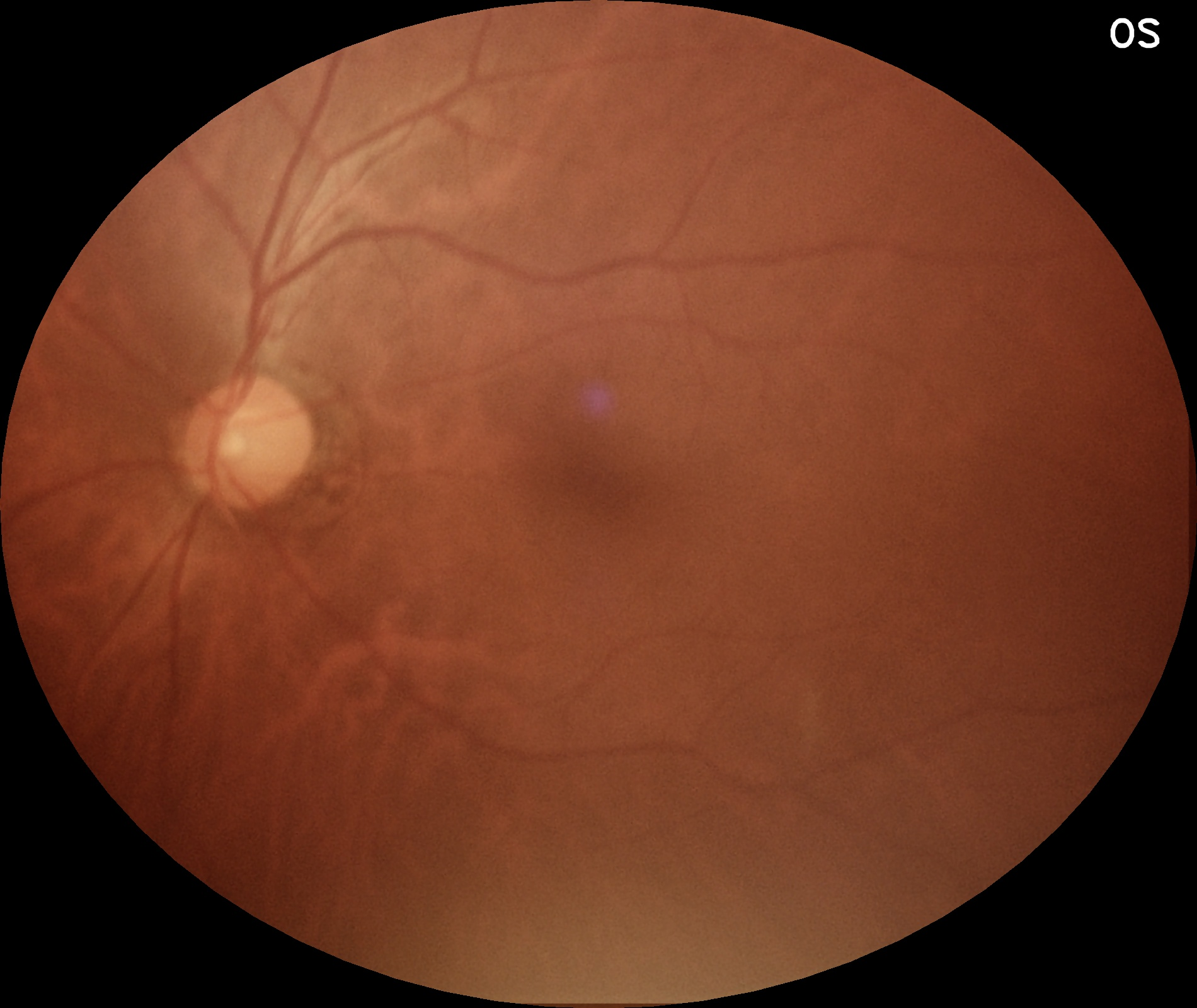
Purpose-built screens for each role, showing real fundus images for the right (OD) and left (OS) eyes.
Reading Doctor screen
Boxes display right and left eye images (multiple images possible). Check boxes below each eye diagnose the patient. A Next button saves the details and loads the next record.
Evaluation Doctor screen
Same image boxes, with the reading doctors’ evaluation labels displayed below. The evaluating doctor then makes their own assessment. The patient status reflects that the evaluating doctor had to make the decision.
Reading doctor evaluations
Doctor A: NPDR Moderate Doctor B: NPDR SeverePower User & Admin
See a summary of images pending across all three doctors in the exam group, and run reports to get the results.